ALGAE CELL ANALYSIS
Consistent label‑free single‑cell measurements – unaffected by autofluorescence or turbidity.
LABEL-FREE CELL ANALYSIS WHERE OPTICAL METHODS REACH THEIR LIMITS
Microalgae are increasingly important in biotechnology for their ability of CO2 sequestration — resulting in applications from biofuel and bioplastics production to nutraceuticals, cosmetics, and wastewater treatment. To enhance circular bioeconomy, monitoring cell viability, concentration, and metabolic state in algal cultures is essential for process optimization and productivity.
Microalgae are autofluorescent due to their photosynthetic pigments (chlorophyll, carotenoids). This autofluorescence directly interferes with fluorescence-based detection methods, complicating staining protocols and reducing measurement accuracy. In addition, dense algal cultures are highly turbid, limiting the effectiveness of optical counting and imaging methods.
Impedance Flow Cytometry (IFC) operates entirely on electrical cell properties and is completely unaffected by autofluorescence or turbidity. This makes it uniquely suited for rapid, label-free characterization of microalgal cultures – providing viability, concentration, and physiological state information with immediate results.
Why Algae Analysis Is Challenging
Unique Properties That Limit Conventional Methods
Microalgae have biological and optical properties that create specific analytical challenges:
01
Autofluorescence Interferes with Optical Detection
Microalgae contain photosynthetic pigments (chlorophyll a/b, carotenoids) that emit strong autofluorescence. This directly overlaps with common fluorescent dyes used in flow cytometry and staining-based viability assays, producing false signals and unreliable results. Additional spectral compensation or specialized dye selection is needed, adding complexity and cost.
Dense Cultures Are Highly Turbid
Productive algal cultures reach high cell densities where the medium becomes opaque. Optical counting methods, image-based analyzers, and OD-based measurements lose accuracy in turbid suspensions. Dilution is often required, introducing variability and additional handling steps.
02
03
Small Cell Size and Morphological Diversity
Microalgae range from approximately 2 to 30 µm and exhibit diverse morphologies – spherical, rod-shaped, flagellated, colonial. This heterogeneity makes standardized optical detection difficult. Cell aggregates and colonial forms further complicate counting and viability determination.
Dynamic Metabolic Changes Are Hard to Track
Algal cells undergo dramatic metabolic shifts depending on light, carbon source, and nutrient availability – such as the transition from photosynthetic to heterotrophic growth, or the accumulation of lipid droplets under stress. These changes affect productivity but are invisible to simple counting or turbidity methods.
04
Comparison of methods for algae analysis
Different analytical methods address different needs. This overview highlights the specific requirements that Impedance Flow Cytometry fulfills.
| Requirement | OD / spectrophotometry Turbidity-based | Hemocytometer Manual / semi-automated | Flow cytometry Fluorescence-based | PAM fluorometry Chlorophyll fluorescence | Amphasys Impedance Flow Cytometry Label-free, impedance-based |
|---|---|---|---|---|---|
| Cell viability | No Turbidity only | Limited Often counted unstained | Impaired Autofluorescence interferes with viability dyes | No Photosynthetic efficiency only | Quantitative Unaffected by pigments |
| Cell count | Indirect Non-linear at high density | Manual / semi-automated Small cells hard to count | Impaired Autofluorescence complicates gating | No | Direct Each cell counted individually |
| Metabolic state | No | No | Limited Autofluorescence complicates markers | Partial Photosynthetic efficiency only | Yes Detects lipid accumulation, stress |
| Works in turbid media | Limited Requires dilution | Limited Visibility issues | Yes | Yes | Yes Impedance is optical-independent |
| Autofluorescence immune | Immune Not fluorescence-based | N/A Often counted unstained | Problematic Spectral overlap with chlorophyll, limited dye choice | Uses it Measures chlorophyll fluorescence | Fully immune Electrical measurement only |
| Time to result | < 1 min | 15–30 min | 30–60 min | Real-time | < 1 min |
| Sample preparation | Dilution + calibration Requires calibration per strain | None or optional staining Often counted unstained | Staining + compensation Limited dye choice due to spectral overlap | None Non-destructive | Dilution + filtration Label-free |
Why IFC Works Where Optical Methods Struggle
The Advantages of Electrical Methods in Autofluorescent Systems
Impedance flow cytometry (IFC) measures the electrical properties of individual algal cells as they pass through a microfluidic channel. IFC analysis relies solely on impedance measurements and not on optical detection. Therefore, it is completely unaffected by autofluorescence, pigmentation, or turbidity of the medium.
The use of electrical signals is thus the key difference when working with photosynthetic organisms. While chlorophyll and phycobiliproteins interfere with all fluorescence-based methods, IFC provides clean and reliable data without spectral compensation or special dye selection.
For algae, this means:
- No interference from chlorophyll or phycobiliprotein autofluorescence
- Accurate measurements in dense, turbid cultures without mandatory dilution
- Simultaneous determination of cell count, viability, and cell state
- Detection of metabolic shifts — including lipid droplet accumulation under stress
- Monitoring of heterotrophic adaptation, which is invisible to PAM fluorometry
- No dyes, no markers, no incubation
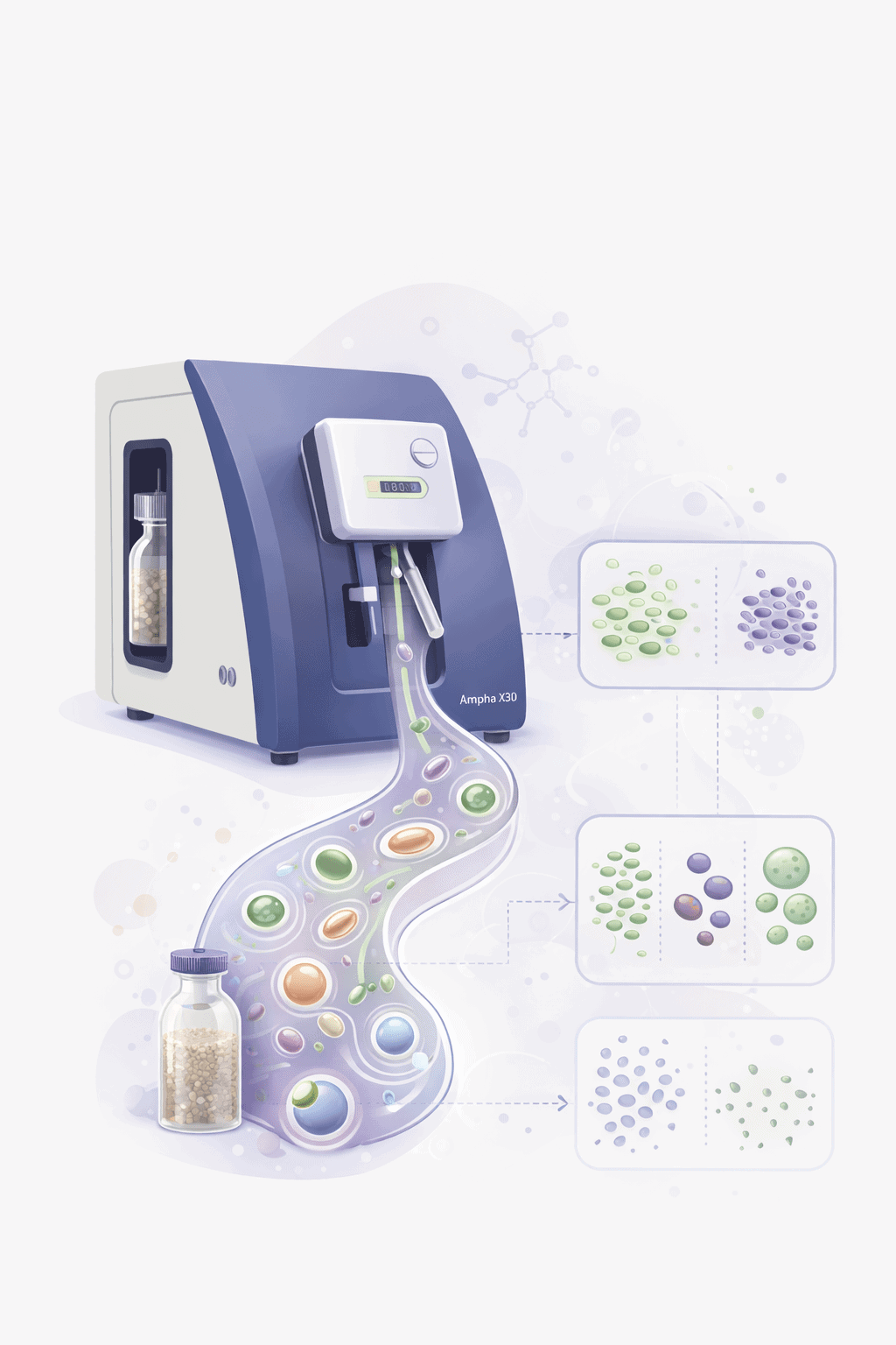
Impedance Flow Cytometry Workflow: From Sample to Decision
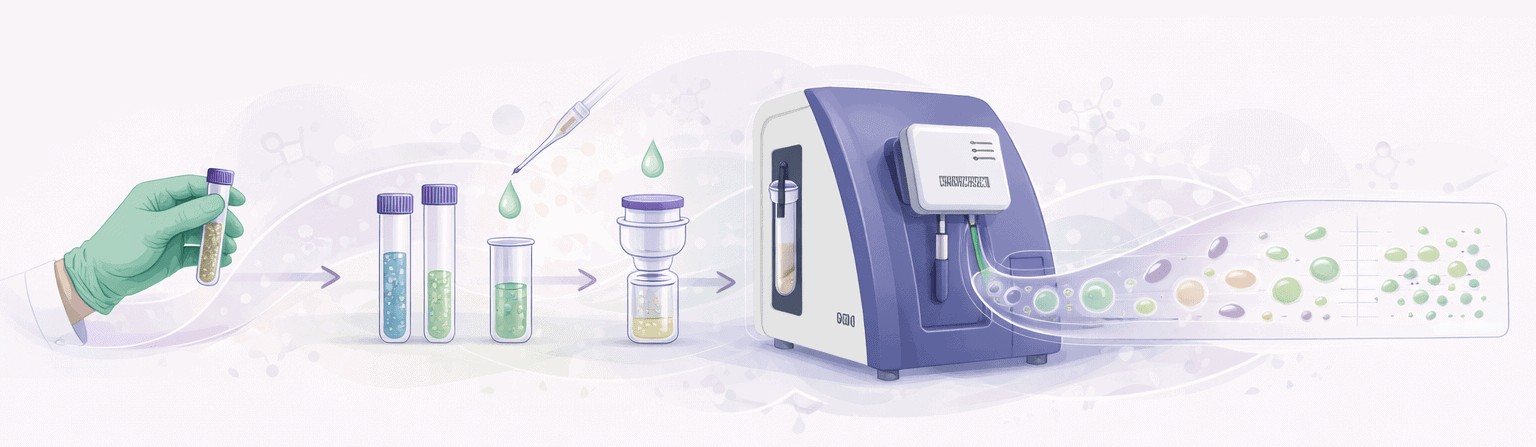
Take a cell sample
Optional dilution
Addition of conductive buffer
Filtration into sampling tube
Load into Ampha X30
Automated measurement
Data analysis + Result
Recommended System for Algae Applications
Quantitative impedance-based algae analysis is performed using:
Ampha X30
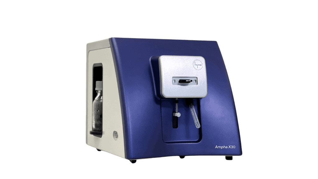
The Ampha X30 is optimized for algae cell analysis and supports:
- Label-free viability, concentration, and metabolic state
- Immune to autofluorescence and turbidity
- Compatible with diverse algae morphologies
- Rapid workflow without dyes or complex sample preparation
- Full flexibility in measurement protocol, gating strategy and data analysis
- Ideal for real-time bioprocess monitoring and cell culture analysis
AmphaChip
The microfluidic AmphaChip is tailored to specific cell sizes to ensure maximum sensitivity.
For algae measurements:
- Channel sizes available for typical microalgae cell sizes (2–30 µm)
- No interference from autofluorescence, debris, or media turbidity
- Measures cell size, membrane capacitance, cytoplasmic conductivity
- Non-invasive measurement
Case Studies
Case Study 1:
Fatty Acid Production in Microalgae
Setup: Microalgae cultures were grown under two conditions: (a) photosynthetic (light, no glucose) and (b) heterotrophic (dark, with glucose). The metabolic adaptation – from green photosynthetic cells to white fatty acid-producing cells – was monitored using impedance flow cytometry.

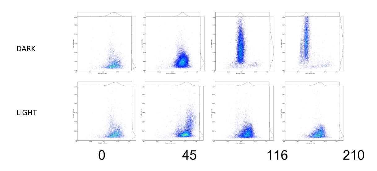
Key Findings: In darkness with glucose, the population grew faster and showed a clear increase in cell volume (amplitude on y-axis) – corresponding to the formation of intracellular fatty acid/lipid droplets. Under photosynthetic conditions, growth was slower and no volume increase was observed. IFC detected these metabolic and morphological changes without labeling or microscopy.
Relevance: Biofuel/biodiesel production, lipid accumulation screening, metabolic engineering, photobioreactor process optimization.
Additional Algae Applications
Photobioreactor Monitoring

Quantitative algal enumeration prior to reactor transfer supports consistent inoculum density and improved batch reproducibility.
Lipid Accumulation Screening

Detection of intracellular volume changes associated with lipid/fatty acid droplet formation under stress conditions.
Strain Comparison

Objective comparison of algal strains under defined stress, light, or nutrient conditions for strain selection.
Stress Response & Contamination

Monitoring of stress-induced physiological changes and detection of contamination events in algal cultures.
Learn More
Deepen Your Knowledge & Drive Better Results
Expert resources on label-free single-cell analysis for algae biotechnology.
Video
Characterisation of Microalgal Cultures by Impedance Flow Cytometry
Prof. Gerhard Obermeyer from the University of Salzburg demonstrates how IFC enables fast, cost-efficient, single-cell insights into algal physiology and viability — using Auxenochlorella protothecoides as a model for scalable algae biotechnology.
Video
Closing the Loop: Automated Real-Time Monitoring
Amphasys CTO Marco Di Berardino presents automated, high-content analysis integrating on-line sampling with label-free single-cell measurements. The principles demonstrated with microbial cultures are directly applicable to algae monitoring.
Video
From Cells to Scale: Navigating the Future of Industrial Bioprocessing
Furkan Gökçe from Amphasys highlights cross-industry challenges in bioprocessing — including automation, advanced monitoring, circular feedstocks, and new infrastructure models relevant to the growing algae biotechnology sector.


Frequently Asked Questions
How does Impedance Flow Cytometry handle autofluorescence in algae?
IFC measures electrical impedance, not optical properties. It is completely unaffected by chlorophyll and phycobiliprotein autofluorescence or any other fluorescent signal. This is the key advantage over fluorescence-based flow cytometry and staining methods for photosynthetic organisms.
Can Impedance Flow Cytometry work with dense, turbid algae cultures?
Yes. Because IFC is based on electrical measurements, media turbidity does not interfere with detection accuracy. For very high-density cultures, a simple dilution step may be needed to ensure proper single-cell passage through the microfluidic channel, but no optical clarity is required.
Which algae species can be measured?
Can Impedance Flow Cytometry detect lipid accumulation in algae?
IFC can detect changes in cell volume and intracellular properties that are associated with lipid droplet accumulation. As shown in the fatty acid production case study, cells accumulating lipids show a measurable increase in impedance amplitude corresponding to increased cell volume. This provides a non-invasive screening parameter for lipid productivity.
What sample preparation is required?
A minimal sample preparation is required: take a sample from the culture, optionally dilute, add conductive measurement buffer, filtrate, and load into the Ampha X30. No staining, no incubation, no calibration.
How does Impedance Flow Cytometry compare to PAM fluorometry?
PAM fluorometry measures photosynthetic efficiency specifically – it provides information about photosystem health but not cell viability, concentration, or overall metabolic state. IFC provides a broader view: viability, cell count, cell size, and physiological state in a single measurement. The two methods are complementary: PAM for photosynthesis-specific questions, IFC for comprehensive cell characterization and process control.
Is Impedance Flow Cytometry suitable for monitoring photobioreactors?
Yes. IFC provides rapid real-time measurements of viability and concentration that support photobioreactor process control. Immediate results enable timely decisions about nutrient feeding, light regime adjustments, or harvest timing.
Talk to a Bioprocessing Expert
Interested in label-free algae analysis without autofluorescence interference? Our application specialists can help you evaluate IFC for your specific algal culture or production process.