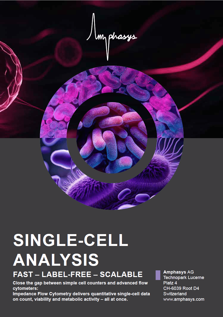
HUMAN AND ANIMAL CELL ANALYSIS
Label-free cell characterization: single-cell resolution in real-time.
LABEL-FREE CELL CHARACTERIZATION FOR CELL CULTURE MONITORING
Human and animal cells analysis is essential in modern biomanufacturing — from monoclonal antibodies and vaccine development to cell therapy, regenerative medicine and cultured meat production. Therefore, monitoring cell viability, concentration, and physiological status during cultivation and production is essential for process understanding and product quality.
Established methods such as trypan blue exclusion, automated image-based counters, and conventional flow cytometry fulfill important functions but also have drawbacks: dye-based methods for viability determination alter the sample and can be toxic, image-based cell counting systems struggle with debris, aggregates and opaque media and rely on optical contrast; conventional flow cytometry requires complex sample preparation and trained operators.
In contrast, Impedance Flow Cytometry (IFC) fills this gap and offers a complementary approach: a rapid, label-free single-cell analysis that provides information on viability, concentration, and cell status without staining, incubation, or complex preparation — thus providing a rapid quality control checkpoint where speed and simplicity are crucial.
Why Human and Animal Cell Analysis Is Demanding
Balancing Speed, Accuracy, and Sample Integrity
Human and animal cells, and specifically mammalian cell cultures, are sensitive, heterogeneous, and dynamically changing. That requires analytical methods that balance sensitivity, speed, and minimal sample disturbance.
01
Sample Integrity vs. Depth of Analysis
In many cases, analytical methods require dyes or fluorescent markers that alter or even destroy the cells. In cell therapy and other high-value manufacturing processes, every single cell counts. A method that provides comprehensive information without altering or damaging the cells therefore offers significant added value.
Speed vs. Complexity
Conventional flow cytometry provides highly detailed information about cell properties. However, this high-resolution data requires complex sample preparation and trained personnel for data analysis. Simpler methods, such as trypan blue staining, are fast but provide only binary information with limited significance. To bridge this gap, there is a need for a rapid, gentle, and informative analytical method. Consequently, there is a need for a rapid, gentle, and informative analytical method.
02
03
Population Heterogeneity
Mammalian cultures are inherently heterogeneous. As a result, cells are in different cycle phases, metabolic states, and stress levels. Bulk measurements only capture averages and do not detect subpopulations with different characteristics. Measurements with single-cell resolution are necessary to identify subpopulations and early process deviations.
Regulatory Challenges
Furthermore, mammalian cell–based products are subject to regulatory oversight requiring that viability, functionality, identity, and purity are demonstrated with accuracy and reproducibility, alongside full software‑based traceability. Analytical methods must be sensitive to heterogeneity, reproducible across operators, non‑perturbing to cells, and validated under GMP (21 CFR Part 11, Annex 11) to ensure compliance throughout the manufacturing process.
04
Comparison of methods for human and animal cell analysis
Different analytical methods address different needs. This overview highlights the specific requirements that impedance flow cytometry fulfills.
| Requirement | Trypan blue Manual / semi-automated | Automated counters Vi-CELL, Countess, etc. | Flow cytometry Fluorescence-based | Capacitance probes In-line, real-time | Amphasys Impedance Flow Cytometry Label-free, single-cell |
|---|---|---|---|---|---|
| Cell viability | Binary Dead / alive | Binary Dye-based | Quantitative With staining markers | Trend Bulk viable biomass | Quantitative Label-free, single-cell |
| Cell count | Manual / semi-automated Hemocytometer | Automated Imaging-based | Direct Each cell counted individually | Trend only Estimated viable biomass | Direct Each cell counted individually |
| Metabolic state | No | No | Yes Dedicated markers needed | No | Yes Membrane + cytoplasm |
| Cell integrity | Altered Dye is cytotoxic | Altered Dye-based | Altered Labels change physiology | Intact Non-invasive | Intact Cells fully reusable |
| Time to result | 5–15 min | 2–5 min | 30–60 min | Real-time | < 1 min |
| Operator dependency | High (manual) Low when automated | Low | High Skilled personnel needed | Low | Low Reproducible results |
| Sample preparation | Staining | Staining + slides | Staining + incubation Complex protocols | Calibration Per cell type and conditions | Optional dilution Label-free |
| Single-cell resolution | No | Limited | Yes | No Bulk signal | Yes Every cell measured |
What IFC Adds to Your Analytical Toolkit
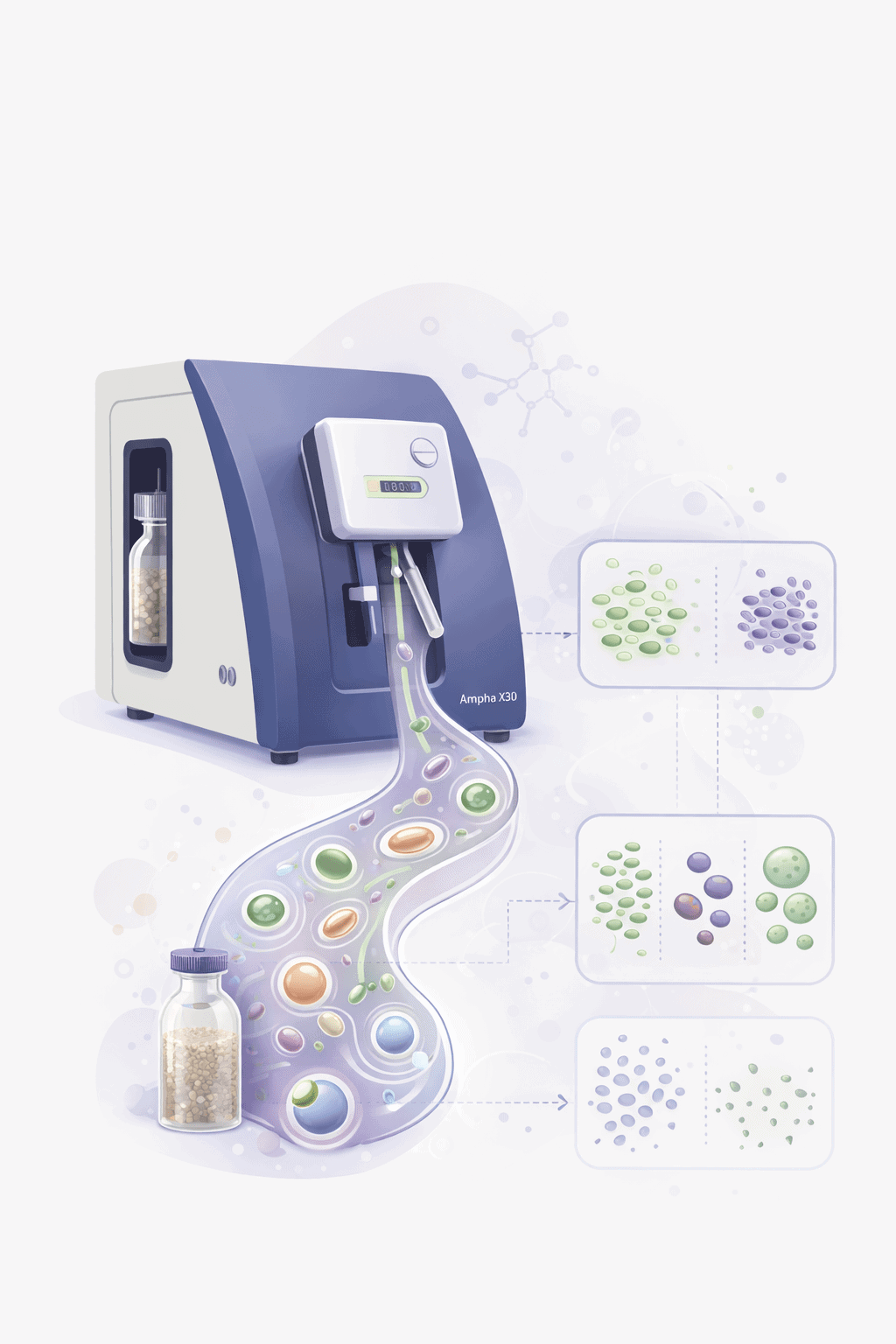
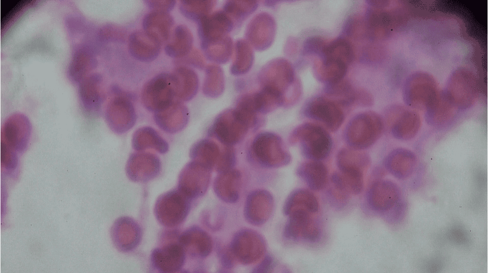
Impedance Flow Cytometry measures the response of individual human and animal cells as they pass through a microfluidic channel with an electrical field. Each cell generates a unique impedance signal — reflecting its size, membrane integrity, and cytoplasmic conductivity without any labels, dyes, or complex preparation.
Among the various methods for human and animal cells analysis, impedance flow cytometry occupies a unique position: it combines the speed and simplicity of counting and viability methods with the single-cell resolution of fluorescence flow cytometry – while preserving the sample completely intact for subsequent use.
IFC stands out from existing methods in the following respect:
- Viability and cell count simultaneously from a single measurement
- Label-free analysis — cells remain unaltered and reusable
- Highly sensitive detection of early metabolic stress based on cytoplasmic conductivity – before viability declines
- Real-time level: minimal sample preparation and immediate results
- No impairement due to turbidity, opacity or particles
- GMP-ready
As a result, Impedance Flow Cytometry is particularly valuable for human and animal cell analysis in process development and continuous cell culture monitoring, for rapid quality control, and for reuse of cells.
Impedance Flow Cytometry Workflow: From Sample to Decision
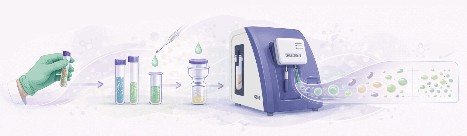
Take a cell sample
Optional dilution
Addition of conductive buffer
Filtration into sampling tube
Load into Ampha X30
Automated measurement
Data analysis + Result
Recommended System for Human and Animal Cell Applications
Quantitative impedance-based analysis for cell culture monitoring:
Ampha X30
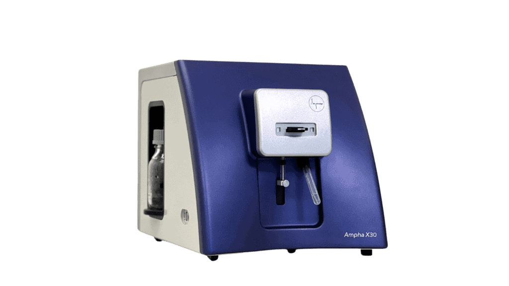
The Ampha X30 is the platform instrument for label-free single cell analysis.
- Determination of viability, concentration, and cell status
- Label-free, minimum sample preparation and immediate results
- Compatible with cell sizes from 1µm to >50 µm
- Reuse of cells for regenerative medicine or cell sorting
- Sophisticated software, GMP-ready
AmphaChip
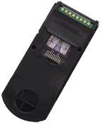
The microfluidic AmphaChip is tailored to specific cell sizes to ensure maximum sensitivity.
- Various channel sizes guarantee for highgest sensitivity
- Determination of cell size, membrane capacitance, cytoplasmic conductivity
- No interference from debris or turbidity
- High accuracy at low concentrations
- Full protocol and gating flexibility
Case Studies
Case Study 1:
Metabolic Status Monitoring of CHO Cells
Three-Week Culture – Viability and Metabolic State
Setup: Adherent CHO cells monitored over 3 weeks: day 4 (blue), day 9 (red), day 19 (green). Viability, count, and metabolic status tracked.
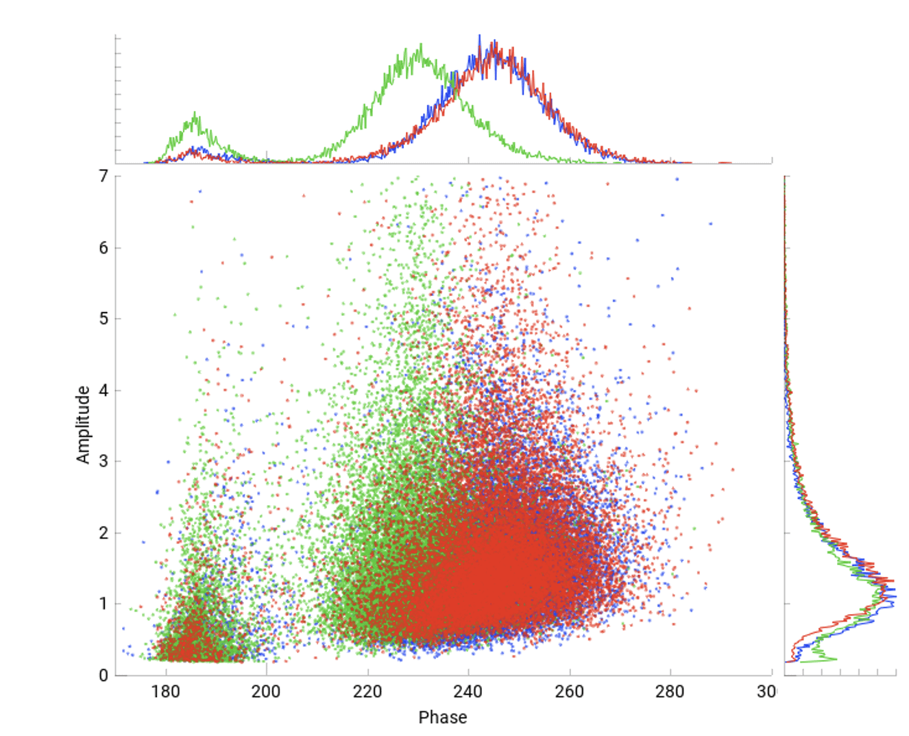
Key Findings: Days 4–9: stable culture conditions, no shift in impedance properties. Day 19: significant shift toward lower phase angles and increase in dead population (see histogram above). The shift indicates that cells are starving due to substrate depletion stress – visible before conventional viability decline.
Relevance: Process development, feeding strategy optimization, early culture decline detection.
Case Study 2:
Apoptosis Detection Without Dedicated Assays
Staurosporine-Induced Apoptosis in BL2 Cells
Setup: Detection of apoptosis requires specific kits in fluorescence flow cytometry. In this example, BL2 cells were treated with apoptosis inducer Staurosporine. Viability was tracked over a period of 60 hours.

Key Findings: The control shows a stable viability over more than 10h. The treated cells, measured without staining or markers and real-time results show a rapid viability drop starting at 1h after induction.
Relevance: Drug screening, cytotoxicity testing, process development.
Additional Applications
Process Development & Scale-Up

Real-time detection of viability and concentration during process development, scale-up and production.
Cell Banking & Thaw Recovery

Rapid assessment of viability for quality control without cell loss through staining.
Bioreactor Monitoring
Rapid, supplementary quality control during the upstream production of biologics, vaccines or recombinant proteins.
Cellular Agriculture

Viability and growth monitoring during expansion of cells for cultivated meat and cell-based food products.
Learn More
Deepen Your Knowledge & Drive Better Results
Expert resources on label-free cell analysis for mammalian bioprocessing.
Video
Navigating the Complexity of Next-Gen Mammalian Bioprocessing
This session explores how emerging technologies address challenges in bispecific antibodies, cell and gene therapies, and cultivated meat. Case studies show how advancements in process analytics drive efficient, cost-effective solutions
Presentation
Manufacturing Processes for Animal Cell-Based Cultured Food
Richard Alldread from The Cultured Hub outlines the technical, economic, and regulatory hurdles in cultured food production — and how adaptable, cost-efficient platforms can accelerate development and scale-up. Slide deck available for download
Download Presentation
Presentation
Monitoring Next-Gen Protein Degrader Bioproduction with IFC
Lorena Jukic from GlycoEra shows how impedance flow cytometry enables precise cell counting and morphology tracking during bioprocess development with Leishmania tarentolae — an unconventional production host. Slide deck available for download.
Download Presentation


Frequently Asked Questions
What cell types can the Ampha X30 measure?
The Ampha X30 supports th emeasurement of all human and animal cells. This includes CHO, HEK293, hybridomas, Vero, insect cells (Sf9, Hi5), primary cells, and stem cells. The sizes are limited by the channel width of 50 µm. However, with a special set-up, small spheroids and organoids can be measured as well. Contact us for your specific application.
How does Impedance Flow Cytometry compare to trypan blue exclusion?
Trypan blue enables a binary classification of dead/alive cells. Impedance flow cytometry (IFC) measures the electrical properties of each individual cell—viability, cell count, and cell state—without dyes or markers. IFC measurement results are not only operator-independent and available within one minute, but the cells also remain unchanged and can be reused.
How does Impedance Flow Cytometry compare to conventional flow cytometry?
Impedance flow cytometry (IFC) bridges the gap between conventional flow cytometry, with its high specificity but complex and time-consuming sample preparation and data analysis, and simpler cell counting and viability determinations. IFC enables rapid, label-free counts as well as viability and cell state measurements with single-cell resolution. Thanks to its real-time results, IFC is the ideal tool for the development, optimization, and scaling of bioprocesses. It fills the gap where speed and simplicity are more important than detailed phenotypic information.
Can Impedance Flow Cytometry detect apoptosis?
Yes. IFC captures membrane integrity and intracellular changes as each individual cell passes through the electrical field of the microfluidic channel. This signal is immediately converted into information: Is a cell dead, alive, starving, or undergoing apoptosis? Neither staining nor specific apoptosis kits are required; apoptosis can be detected directly as it occurs.
What sample preparation is required?
Minimal sample preparation is required. Depending on the concentration, the samples are diluted, then mixed with a conductive buffer, filtered, and loaded directly into the instrument. Staining, labeling, or incubation is unnecessary. Less than 5 minutes elapse from sampling to result.
What is the measurement throughput?
The measurement itself takes 30–60 seconds. Sample preparation depends on the total cell concentration and may require a dilution step. A measurement, including sample preparation and data analysis, typically takes less than 5 minutes.
Talk to a Bioprocessing Expert
Ready to improve your cell culture monitoring with real-time, label-free cell analysis? Our experts are here to help you find the right solution for your specific application.