
Technology
Impedance Flow Cytometry for Real-Time Cell Culture Monitoring
Real-Time Single-Cell Data for Smarter Decisions
Cell viability

Know your true viable fraction
Real measured viability of each single cell. No indirect measurement or correlation. Immediate results without dyes or markers. And the dead cells are counted exactly as well.
Cell count

Absolute numbers, not estimates
Direct counting of each individual cell, regardless of its size. No indirect determination, no calibration curves; works even in turbid, opaque, and autofluorescent media.
Cell state

Are your cells healthy?
The Measurement Principle: Impedance Flow Cytometry
Impedance flow cytometry is an analytical technique based on the characterization of individual cells flowing through a microfluidic channel under the influence of an applied alternating current field. The resistance (impedance) of the cells in the electric field provides a specific signal that reflects their state.
Single-Cell Flow through Microfluidic Chips
The cells, suspended in a conductive buffer, flow through the channel of a microfluidic chip. Hydrodynamic focusing ensures that the cells pass through the measurement zone individually. Thanks to the channel size being adapted to the cell size, high sensitivity and reproducibility of the measurements are ensured across different cell types and media conditions.
Electrical Impedance Measurement
Every cell passing through the electrodes is influenced by the applied alternating electric field. The simultaneous application of multiple frequencies stimulates different cell properties. The impedance signal depends on:
- Cell size and volume (cell count)
- Membrane capacitance (viable/dead cells)
- Cytoplasmic conductivity (cell metabolism and health state)
Consequently, by distinguishing between membrane-bound and cytoplasmic properties, multifrequency measurements provide a wealth of information in real time.

Population-Level Data Generation
See the Measurement Principle in Action
Understanding how impedance flow cytometry works explains why it delivers objective and reproducible results—regardless of cell type, media turbidity, or user experience.
In particular, this short explanatory video illustrates how the system electrically characterizes individual cells in a precisely defined microfluidic channel.
What Bioprocessing Technology Measures — and Why It Matters
Impedance measurement data provides biophysical information that allows conclusions to be drawn about various cell properties and serves as a basis for decision-making.

Membrane Integrity
Membrane integrity corresponds to cell viability. It allows the differentiation of viable cells with intact membranes from damaged or dead cells – the most fundamental parameter for the health of a cell culture.

Cytoplasmic Conductivity
Cytoplasmic conductivity is a measure of intracellular composition and metabolic state. Changes in conductivity can be associated with stress (e.g., starvation), metabolic changes (e.g., intracellular lipid synthesis), or adaptation to culture conditions.

Cell Size and Volume
The impedance signal provides information about cell size and volume at every frequency. Shifts in the signal’s magnitude reflect changes in cell properties that occur independently of other measurement results.

Population Heterogeneity
Cell populations are highly heterogeneous and exist in parallel in different states. While bulk measurements only provide average values, impedance flow cytometry enables the unambiguous identification and quantification of these subpopulations.

Cell Concentration
Each cell passing through the electric field generates a signal that triggers cell counting. This enables the precise counting of low cell concentrations and cells of small size, as detection is non-optical. Turbidity, cell debris, or autofluorescence pose also no problem.
From sample to decision in under 5 minutes
Minimal preparation. No staining, no markers. No incubation. No calibration.
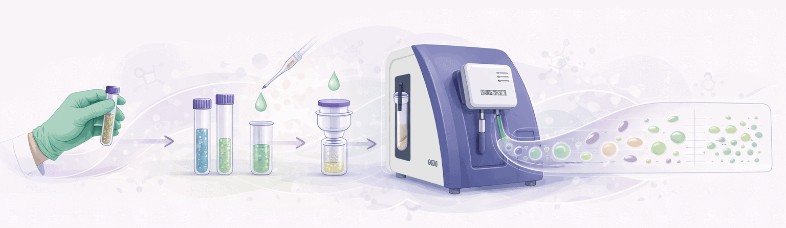
Take a cell sample
Optional dilution
Addition of conductive buffer
Filtration into sampling tube
Load into Ampha X30
Automated measurement
Data analysis + Result
Comparison of methods for cell analysis
Different analytical methods address different needs. This overview highlights the specific requirements that impedance flow cytometry fulfills.
| Requirement | OD600 Spectrophotometry | CFU plating Colony counting | Dye-based staining Trypan blue, methylene blue | Flow cytometry Fluorescence-based | Capacitance probes In-line, real-time | Amphasys Impedance Flow Cytometry Label-free, single-cell |
|---|---|---|---|---|---|---|
| Cell viability | No Turbidity only | Culturable only Misses VBNC cells | Binary Dead / viable | Quantitative With appropriate dyes | Trend Bulk viable biomass | Quantitative Label-free, single-cell |
| Cell count | Indirect Arbitrary units | Yes After 24–72 h | Manual / semi-automated Hemocytometer | Direct Each cell counted individually | Trend only Estimated viable biomass | Direct Each cell counted individually |
| Metabolic state | No | No | No | Yes Dedicated markers needed | No | Yes Membrane + cytoplasm |
| Cell integrity | Intact | Intact Plated on agar | Altered Dye is cytotoxic | Altered Labels change cells | Intact Non-invasive | Intact Cells fully reusable |
| Time to result | < 1 min | 24–72 hours | 5–30 min | 30–60 min | Real-time | < 1 min |
| Operator dependency | Low | High | High (manual) Low when automated | High Skilled personnel needed | Low | Low Reproducible results |
| Sample preparation | Dilution + calibration | Dilution + plating + incubation | Staining + counting | Staining + incubation | Calibration Per organism and conditions | Minimal Optional dilution and filtration, label-free |
| Single-cell resolution | No Bulk signal | No Colony level | Limited | Yes | No Bulk signal | Yes Every cell measured |
| Works in turbid / autofluorescent media | Limited Affected by turbidity | Yes Culture-based | Limited Optical interference | Limited Autofluorescence issues | Yes | Yes Impedance is optical-independent |
Bioprocessing Technology Designed for Your Workflow
One platform. Three components. Tailored to your cell type and workflow.
Ampha X30
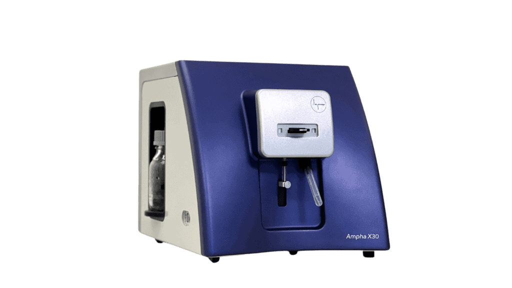
Text: The cell analyzer for real-time, label-free determination of viability, concentration, and cell state. From sample to result in under 5 minutes — across bacteria, yeast, mammalian cells, and algae.
AmphaChip
Microfluidic chips in multiple channel sizes — optimized for your cell type. Simply switch the chip to move between organisms. No optical adjustments, no recalibration.
AmphaSoft

Purpose-built analysis software for gating, visualization, batch comparison, and data export. Available in Standard and Pro editions — designed for both routine monitoring and in-depth analysis.


Frequently Asked Questions
How does impedance flow cytometry differ from fluorescence-based flow cytometry?
Conventional flow cytometry relies on application- and cell-specific markers and optical detection. As a result, this requires complex and time-consuming sample preparation and trained personnel for data analysis. Impedance flow cytometry, on the other hand, is based on the label-free measurement of electrical cell properties, thus requiring virtually no sample preparation and delivering results in real time.
What cell types can be analyzed?
Amphasys cell analyzers utilize microfluidic chips with varying channel diameters, which are tailored to cell size for maximum sensitivity. This allows for the measurement of the entire spectrum of cells, from bacteria, algae, and yeast to human and animal cells down to a size of approximately 50 µm.
For larger cells, spheroids, and organoids, please contact us.
Does IFC measure viability?
Yes. Cells can be viable but – for whatever reason – fail to divide. In contrast, while CFU counting only counts and records culturable cells, IFC allows for the precise quantification and differentiation of viable and dead cells.
Can the system detect VBNC (viable but non-culturable) cells?
Yes. Unlike CFU plating, which only detects cells capable of forming colonies, IFC detects cells based on their biophysical properties. Specifically, VBNC cells with intact membranes are measured and classified as viable — providing a more complete picture of population status.
Is the measurement affected by media turbidity or autofluorescence?
No. Because IFC is based on an electrical signal rather than optical detection and offers high sensitivity, it is inherently insensitive to turbidity, opacity, or autofluorescence. As a result, this is a significant advantage over optical methods and allows even the measurement of low concentrations or small cells under challenging conditions.
How does Impedance Flow Cytometry compare to OD600?
Optical density measurement at 600 nm (OD600) correlates the loss of transmitted light with cell concentration. Furthermore, it cannot distinguish between viable and dead cells, nor can it determine cell size or biomass composition. OD600 measurements are affected by the medium, therefore a blank measurement is required for calibration. Impedance flow cytometry (IFC) allows for direct single-cell counting with simultaneous determination of cell viability, requires neither labeling nor incubation or calibration, and is independent of turbidity.
How long does a measurement take?
A complete measurement, including sample preparation and measurement, takes approximately 2–3 minutes. The entire workflow, from sample collection to result, typically takes less than 5 minutes. The system can process multiple samples sequentially.
Ready to See IFC in Action?
Contact our application team to discuss how impedance flow cytometry fits your bioprocess monitoring — or request a measurement with your own samples.